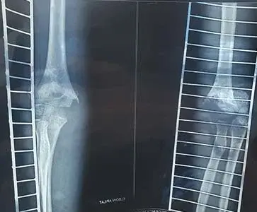
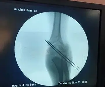
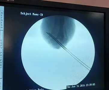
Supracondylar Fractures are one of the most common traumatic fractures seen in children and most commonly occur in children 5-7 years of age from a fall on an outstretched hand.
Diagnosis can be made with plain radiographs.
Treatment is usually closed reduction and percutanous pinning (CRPP), with the urgency depending on presence or absence of hand perfusion.
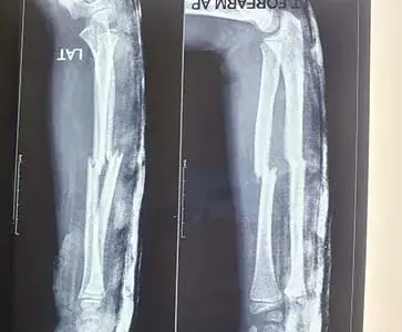
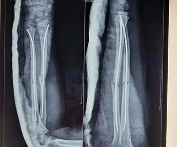
The forearm is the part of the arm between the wrist and the elbow. It is made up of two bones: the radius and the ulna. Forearm fractures are common in childhood, accounting for more than 40% of all childhood fractures. About 3 out of 4 (75% of) forearm fractures in children occur at the wrist end of the radius.
Forearm fractures often occur when children are playing on the playground or participating in sports. If a child takes a tumble and falls onto an outstretched arm, there is a chance it may result in a forearm fracture. A child's bones heal more quickly than an adult's, so it is important to treat a fracture promptly—before healing begins—to avoid future problems.

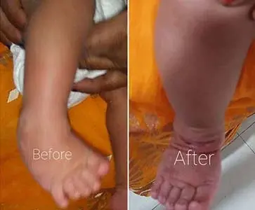
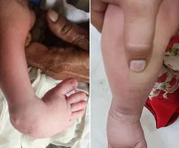
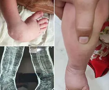
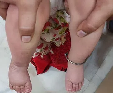
Most commonly, a doctor recognizes clubfoot soon after birth just from looking at the shape and positioning of the newborn's foot. Occasionally, the doctor may request X-rays to fully understand how severe the clubfoot is, but usually X-rays are not necessary.
It's possible to clearly see most cases of clubfoot before birth during a routine ultrasound exam in week 20 of pregnancy. While nothing can be done before birth to solve the problem, knowing about the condition may give you time to learn more about clubfoot and get in touch with appropriate health experts, such as a pediatric orthopedic surgeon and a genetics counselor.
Because your newborn's bones, joints and tendons are very flexible, treatment for clubfoot usually begins in the first week or two after birth. The goal of treatment is to improve the way your child's foot looks and works before he or she learns to walk, in hopes of preventing long-term disabilities.
Treatment options include:
