×


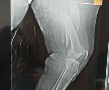
Distal femur fractures are traumatic injuries involving the region extending from the distal metaphyseal-diaphyseal junction to the articular surface of the femoral condyles.
Diagnosis is made radiographically with CT studies often required to assess for intra-articular extension.
Treatment is generally operative with ORIF, intramedullary nail, or distal femur replacement depending on available bone stock, age of patient, and patient activity demands.


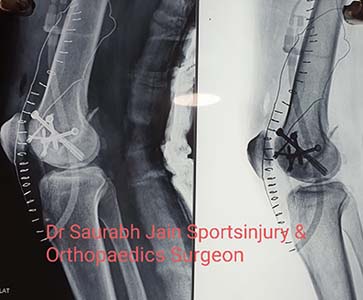
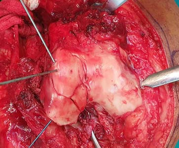
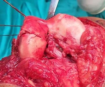
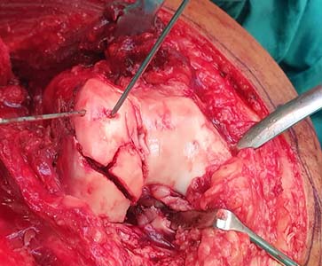
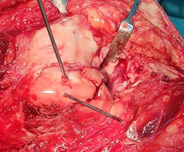
Anatomic reduction of the articular surface, stable fixation, and early mobilization should be the aims of treatment. Open reduction and internal fixation is the 1st choice for the treatment of displaced Hoffa fractures, and it is also suitable for the treatment of nondisplaced Hoffa fractures. The knee joint is placed in flexion during surgery, placing the joint capsule and gastrocnemius in a relaxed state, which reduces the traction on the fracture and is conducive to fracture repair. The appropriate surgical plan is chosen based on the location of the Hoffa fracture, characteristics of the fracture line, fracture severity, and associated injuries.


These fractures occur at the ankle end of the tibia. They are also called tibial plafond fractures. One of the common types in children is the distal tibial metaphyseal fracture. This is a fracture in the metaphysis, the part of tibia before it reaches its widest point.
These fractures are usually transverse (across) or oblique (slanted) breaks in the bone. Distal tibial metaphyseal fractures usually heal well after setting them without surgery and applying a cast. However, there is a risk of full or partial early closure of the growth plate. This may lead to a growth arrest in the form of leg length discrepancy or other deformity.


The proximal tibia is the upper part of the shinbone that connects to the knee joint. Proximal tibia fractures are fairly common lower-leg injuries. They can result from low-energy injuries or a high-energy injury, ranging from slips and falls to major car accidents.
Because blood vessels, ligaments, muscles, nerves and skin may be injured simultaneously during this type of fracture, it is crucial that an orthopedic specialist assesses any damage to soft tissue to properly treat the facture. Proximal tibia fractures must be properly identified and diagnosed to manage the injury and restore normal range of motion, stability and strength to the limbs. Proper, simultaneous management of such fractures and any accompanying soft tissue injuries can also reduce the likelihood of arthritis in the future.


The most common treatment for intertrochanteric fractures is surgery. In most cases, surgery is recommended because this fracture can take a long time to heal on its own. One of the most common surgical treatments for this type of hip fracture is an open reduction and internal fixation (ORIF). This is a type of surgery that puts the broken bone in place and fixes it with screws, rods, pins, or plates.
However, surgery may not be an option if you have bleeding problems or can’t tolerate anesthesia.


Patella Fractures are traumatic knee injuries caused by direct trauma or rapid contracture of the quadriceps with a flexed knee that can lead to loss of the extensor mechanism.
Diagnosis can be made clinically with the inability to perform a straight leg raise and confirmed with radiographs of the knee.
Treatment is either immobilization or surgical fixation depending on fracture displacement and integrity of the extensor mechanism.


The tibia or shin bone is a major bone of the leg which connects the knee to the ankle. A tibial fracture is a break in the continuity of the shin bone (tibia).
Fractures of proximal tibia: A proximal tibial fracture is a break in the upper part of the shin bone or tibia. Proximal tibial fractures may or may not involve the knee joint. Fractures that enter the knee joint may cause joint imperfections, irregular joint surfaces, and improper alignment in the legs. This can lead to as joint instability, arthritis, and loss of motion. These fractures are caused by stress or trauma or in a bone already compromised by disease, such as cancer or infection. Proximal tibia fractures can result in injury to the surrounding soft tissues including skin, muscle, nerves, blood vessels, and ligaments.
The symptoms of tibia fracture include painful weight bearing movements, tenseness around the knee, limitation of movement and deformity around the knee. In some individuals, impairment of blood supply secondary to the fracture may result in a pale or cool foot. Patients may also experience numbness or feelings of ‘pins and needles’ in the foot as a result of associated nerve injury.
The management of the fracture is based on the severity of the fracture, medical condition of the patient and the patient’s lifestyle.
Non-surgical treatment comprises of immobilizing the fractured site with the help of casts or braces to prevent weight bearing and to help the healing process. X-rays are taken at regular intervals to assess the healing process. Weight bearing and movement are initiated gradually, depending on the nature of the injury and the condition of the patient.
Surgical treatment is considered to maintain alignment of the fractured bone. External or internal fixators may be used to align the fractured bone segments. If the fracture does not involve the knee joint, rods and plates can be used to stabilize the fracture. For a fracture involving the knee joint, a bone graft may be required to prevent the knee joint from collapsing. An external fixator is used when the surrounding soft tissue is severely damaged, as the use of plate and screw may be harmful.
As the tibial fracture usually involves the weight bearing joint it may cause long term problems such as loss of knee motion or instability and long term arthritis. Hence a rehabilitation program is initiated along with the treatment comprising of instructions on weight bearing, knee movements, and the use of external devices such as braces


A fracture, or break, in the shinbone just below the knee is called a proximal tibia fracture. The proximal tibia is the upper portion of the bone where it widens to help form the knee joint.


Pubic symphysis diastasis injuries typically occur following a high velocity force such as road traffic accidents, horse riding, crush injuries and falls from a height. There are reports of a range of injuries caused by waterslides including spinal fractures, head injuries and vaginal injuries. Here we report the case of a 50-year-old female who suffered a pelvic diastasis injury during the use of a waterslide.


This is done with a tendon taken from another place in your foot. In some cases, the Achilles tendon repair surgery can be done as a minimally invasive procedure. This is done with several small incisions instead of one large one. It may use a special scope with a tiny camera and a light to help do the repair.


Intramedullary nailing is the most popular and widely used method for treating tibial shaft fractures. Intramedullary nailing involves minimal surgical dissection, allowing preservation of blood supply by not disrupting the soft tissue around the fracture.