Joint pain from cartilage damage can make daily activities like walking, climbing stairs, or exercising feel overwhelming.Micro drilling surgery offers a cutting-edge, minimally invasive solution to repair cartilage and restore joint function, helping patients return to their active lives with ease. Dr. Saurabh Jain, a leading arthroscopic and sports injury surgeon , provides expert care tailored to individual needs, using advanced techniques like keyhole bone drilling treatment to promote healing. Whether you’re an athlete, student, or office worker, this procedure can help you regain mobility and comfort.
What Is Micro Drilling Surgery?
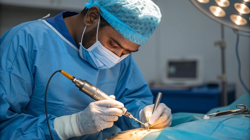
Micro drilling surgery, also known as microfracture surgery, is a minimally invasive procedure to treat cartilage damage in joints, most commonly the knee, but also applicable to the ankle, hip, or shoulder. It involves creating small holes in the subchondral bone beneath damaged cartilage to stimulate the release of bone marrow, which forms a blood clot that promotes new cartilage growth. This arthroscopic micro drilling procedure uses a camera and specialized tools inserted through tiny incisions, ensuring precision and reduced tissue trauma. The procedure is ideal for small cartilage defects (less than 2-3 cm²) caused by trauma or overuse, offering a less invasive alternative to joint replacement. It typically takes 30-60 minutes under anesthesia, with patients often discharged the same day. The subchondral drilling technique leverages the body’s natural healing process, making it a cornerstone of cartilage repair for active individuals.
Causes and Symptoms of Cartilage Damage
Cartilage damage occurs when the smooth, cushioning tissue in joints wears down or is injured, leading to pain and reduced mobility. Common causes include acute trauma from falls or sports injuries, repetitive stress from overuse, or degenerative conditions like early osteoarthritis. Athletes are at higher risk due to repetitive impact, while office workers may develop issues from prolonged sitting or poor posture. Aging also contributes, as cartilage loses its elasticity over time. Symptoms vary depending on the damage’s severity. Patients often experience localized joint pain, especially during movement, accompanied by swelling or stiffness. A grinding or catching sensation may occur, and in severe cases, the joint may lock or give way. If untreated, cartilage damage can lead to chronic pain or arthritis, making early intervention critical.
Common Causes
- Trauma: Falls, collisions, or direct blows to the joint.
- Overuse: Repetitive movements in sports or daily activities.
- Degeneration: Age-related wear or early osteoarthritis.
Recognizing Symptoms
- Pain: Localized discomfort, worse with activity.
- Swelling and Stiffness: Fluid buildup or reduced range of motion.
- Joint Instability: Catching, locking, or giving way.
Prompt recognition ensures timely treatment to prevent further damage.
Diagnosis of Cartilage Damage
Diagnosing cartilage damage involves a combination of clinical evaluation and imaging to assess the extent of injury. The process begins with a detailed history, discussing the injury’s mechanism, such as a fall or repetitive stress. Physical exams evaluate joint tenderness, range of motion, and stability, with tests like the McMurray test for knee cartilage damage.
Imaging confirms the diagnosis, with MRI being the gold standard for visualizing cartilage and soft tissues, offering 90% accuracy. Arthroscopy may be used diagnostically to directly view the joint surface. X-rays help rule out fractures or bony abnormalities. Accurate diagnosis distinguishes cartilage damage from other issues like ligament tears, guiding effective treatment.
Physical Examination
Physical exams focus on joint function and symptoms. Palpation identifies tender areas, while range of motion tests reveal stiffness or locking. Specific maneuvers assess joint stability, helping pinpoint cartilage damage. These non-invasive tests provide immediate insights for treatment planning.
Imaging Tests
MRI: Visualizes cartilage and soft tissue damage.
X-ray: Rules out fractures or bony issues.
Diagnostic Arthroscopy: Directly inspects the joint surface.
These tools ensure a precise diagnosis for tailored treatment.
Treatment Options for Cartilage Damage
Treatment depends on the defect’s size, location, and patient activity level. Non-surgical options suit smaller defects or less active individuals, while pin-hole drilling surgery is recommended for active patients with significant damage. The goal is to restore joint function and prevent arthritis progression.
Non-Surgical Treatments
Non-surgical approaches focus on symptom relief and joint support. Physical therapy strengthens surrounding muscles, improving stability and reducing stress on the cartilage. Bracing or orthotics may support the joint during activities, while rest and activity modification prevent further damage. These methods are effective for small defects or early-stage issues, with 50-60% of patients experiencing symptom improvement.
Surgical Treatments
For larger defects or active patients, micro drilling surgery is the preferred choice. This small-drill joint repair technique creates tiny holes in the subchondral bone to stimulate cartilage regeneration through bone marrow release. Performed arthroscopically, it addresses associated injuries like meniscus tears in the same procedure. The osteochondral micro drilling procedure is highly effective for defects under 2-3 cm², offering a durable solution for joint health.
The Micro Drilling Surgery Procedure
The procedure begins with thorough preparation to ensure optimal outcomes. Pre-operative evaluations include imaging and physical therapy to strengthen the joint. During surgery, small incisions allow the arthroscope and drill to access the joint. Tiny holes are made in the subchondral bone, promoting marrow release to form new cartilage. The tiny hole bone healing surgery approach minimizes trauma, with patients often discharged the same day.
Post-operative care focuses on protecting the joint while encouraging healing. Crutches and bracing are used initially, with early physical therapy to restore mobility.
Pre-Operative Preparation
Preparation includes pre-op physical therapy to enhance muscle strength, reducing recovery time. Patients are advised to optimize nutrition for healing, and anesthesia options (general or regional) are discussed. Medical history is reviewed to minimize risks.
The Surgery Step by Step
- Anesthesia: Administered for comfort, typically general or regional.
- Arthroscopy: Small incisions for camera and drill insertion.
- Drilling: Tiny holes created in subchondral bone to stimulate marrow.
- Closure: Incisions sutured, joint bandaged.
This arthroscopic bone marrow stimulation technique ensures precision and minimal disruption.
Post-Operative Care
Immediate care includes swelling control with ice and elevation. Crutches protect the joint for 6-8 weeks, and a brace may be used. Wound care prevents infection, and follow-ups monitor healing. Early therapy restores range of motion, setting the stage for rehabilitation.
Benefits of Micro Drilling Surgery
Micro drilling surgery offers significant advantages for cartilage repair. The minimally invasive bone drilling approach reduces pain, scarring, and infection risk, with patients often resuming light activities within weeks. The procedure promotes natural cartilage regeneration, delaying or preventing the need for joint replacement. Success rates range from 75-85% for small defects, with improved joint function and reduced pain.
Patients benefit from long-term joint health, enabling active lifestyles without chronic discomfort. The advanced tiny-drill treatment for joints allows precise targeting of damaged areas, enhancing outcomes for athletes or workers.
Recovery and Rehabilitation After Surgery
Recovery is a phased process, starting with joint protection and swelling reduction. Crutches are used for 6-8 weeks to avoid weight-bearing, particularly for knee procedures. Physical therapy begins early, focusing on range of motion, followed by strength and balance exercises. Full recovery takes 6-12 months, with return to high-impact activities at 9 months for most.
Rehabilitation includes quad strengthening, balance training, and gradual activity progression. Consistency is crucial to protect the new cartilage and ensure lasting results.
Immediate Post-Surgery Phase
The first 6-8 weeks focus on rest, ice, and elevation to manage swelling. Crutches and bracing protect the joint, while gentle movements prevent stiffness. Pain is managed with prescribed methods, and wound care minimizes infection risk.
Physical Therapy and Rehab
Therapy starts with passive motion exercises, progressing to strength training for surrounding muscles. Balance tools enhance proprioception, and later phases include low-impact activities like swimming. Sport-specific drills prepare athletes for return.
Timeline for Return to Activities
- 1-2 Weeks: Reduced swelling, gentle motion exercises.
- 6-8 Weeks: Partial weight-bearing, improved range.
- 3-6 Months: Light activities, strength gains.
- 9-12 Months: Full return to high-impact activities.
This timeline ensures safe recovery.
Risks and Complications
Risks include incomplete cartilage healing (15-25% of cases), persistent pain, or scar tissue formation. Infection or blood clots are rare but possible. Overloading the joint too soon can compromise results. Choosing an experienced surgeon and adhering to rehab protocols minimizes these risks.
Micro drilling surgery is a transformative solution for cartilage damage, offering cartilage regeneration and joint health through osteochondral micro drilling procedure. This minimally invasive approach ensures faster recovery and lasting results, ideal for athletes, students, or workers seeking to regain mobility. With expert care, patients can return to their active lives with confidence.
Dr. Saurabh Jain, Arthroscopic and Sports Injury Surgeon in Lucknow